 “A Deep Learning Approach for Osteoporosis Identification using
Cone-beam Computed Tomography (OSTAK)”
Project No. lzp-2021/1-0031

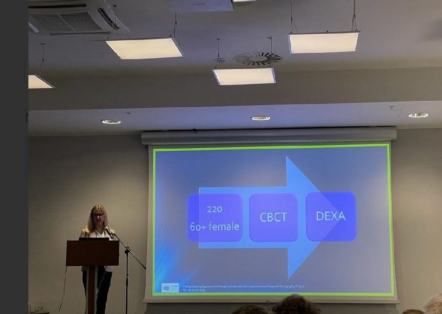
The goal of the interdisciplinary project is to develop an innovative method for the identification the risk of osteoporosis by using Cone-beam Computed Tomography (CBCT) of the maxillofacial region and to evaluate its efficacy by using an end-to-end Deep Learning (DL) approach. CBCT examination is a non-invasive x-ray technology which produces 3D images. Using a DL approach, a Computer Vision method will be elaborated which can identify more quickly and accurately the risk of osteoporosis in women. Consequently, it facilitates the early treatment of the disease as well as prevents osteoporotic fractures. The project will aim for the expansion of personalized medicine, medical and ICT sectors. The project will be conducted by the medical experts from the Rīga Stradiņš University (RSU) and DL researchers from the Institute of Electronics and Computer Sciences (EDI). The patient`s dataset will be collected by RSU researchers, in which CBCT and osteodensitometry studies are planned for 220 patients. The various measurements of the quality and quantity of the bone and radiological bone density in the maxilla and cervical vertebrae will be performed. The results will be used by EDI to develop a computer-based method of semantic segmentation, classification and explainability of osteoporosis.
News Update
01.02.2022 – 30.03.2022
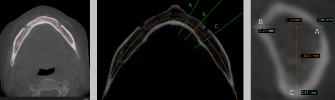
The research strategy, methodology, and tasks for identifying osteoporosis risk factors are being refined. Based on the innovative methodology developed by Riga Stradiņš University (RSU), a protocol for mandibular measurements and indices is being designed by analyzing 3D CBCT images. For protocol validation, seminars are scheduled on 21.02.2023 and 09.05.2023. The Institute of Electronics and Computer Science (EDI) receives initial patient files in .nrrd format and identifies the most suitable Deep Neural Network (DNN) architecture. The ResNet-101 deep learning architecture is selected as the most appropriate.
01.04.2022. – 30.06.2023.
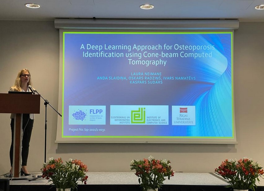
A patient annotation methodology has been developed, along with the presentation of guidelines and software for annotating 3D CBCT images. The annotation process has been initiated. The first research results were presented on June 3–4, 2022, in Riga at the 16th Riga-Rostock International Symposium and the 10th Baltic Association for Maxillofacial and Plastic Surgery Congress titled “Latest Technologies in Oral, Facial, and Maxillofacial Surgery.” Dr. L. Neimane delivered a presentation on “Osteoporosis Detection in Cone-Beam Computed Tomography Examinations Using Deep Machine Learning.”
01.07.2022. – 30.09.2022.
Image annotation continues at RSU. EDI proceeds with the development of software for a modular deep neural network. This network is tested for the classification task, specifically for selecting cross-sections of the mandible where the mental foramen is best visible.
01.10.2022. – 31.12.2022.
Research findings are presented (as posters) and published in the proceedings of the ESHNR 2022 conference on 10.11.2022. The following publications were included:
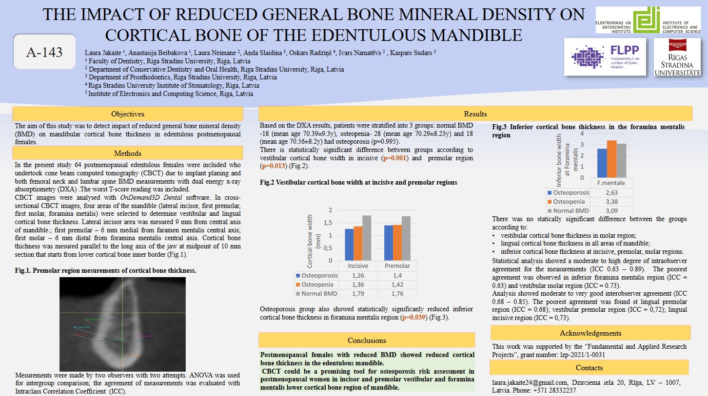
- L. Jakaite et al., “The Impact of Reduced General Bone Mineral Density on Cortical Bone of the Edentulous Mandible”
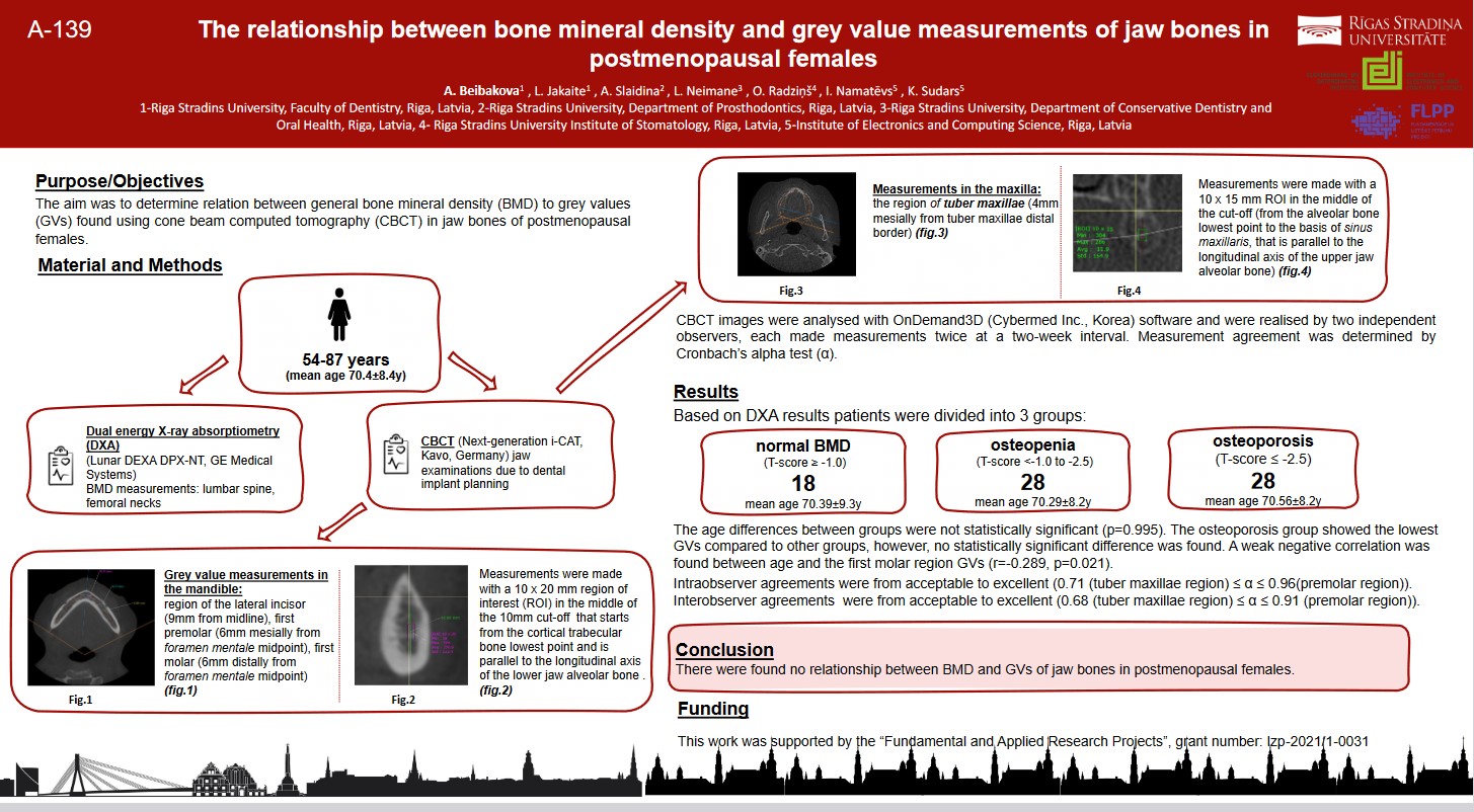
- A. Beibakova et al., “The Relationship Between Bone Mineral Density and Grey Value Measurements of Jaw Bones in Postmenopausal Females.”
01.01.2023. – 31.03.2023.
The annotation of CBCT images has been completed, resulting in the creation of the OSTAK dataset. This dataset includes annotated mandibular images from 188 patients for modular deep neural network training and validation. The data input and software for network training have also been finalized.
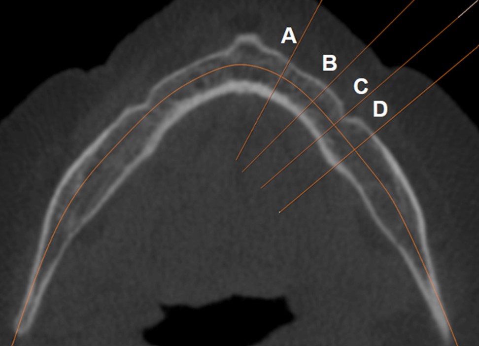
A modular deep neural network based on the ResNet-101 architecture has been trained and validated. The network comprises three sequential deep-learning models for classification tasks and two for regression tasks. The first model identifies the optimal cross-section of the mandible in axial CBCT images where the mental foramen is best visible. The training accuracy reached 98.85% (39 epochs), and validation accuracy reached 93.99% (35 epochs). The second model generates cross-sections of the mandible based on predefined distances, producing images of mandibular cross-sectional areas. The third model measures the cortical bone thickness of the mandible.
25.03.2023.
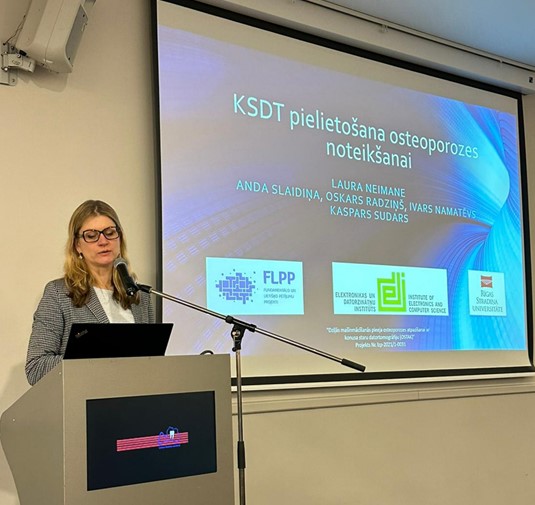
During the Latvian Dental Association meeting, Dr. L. Neimane presented a report on identifying osteoporosis risk in dental, facial, and maxillofacial radiological images. The professor emphasized key aspects that dentists should observe in daily practice to detect osteoporosis risk in postmenopausal women:
Read more on the Latvian Dental Association website.
01.04.2023. – 30.06.2023.
The obtained results have been analyzed, and the software configuration has been fine-tuned. A research paper titled “Modular Deep Neural Network for Detection of Osteoporotic Changes in Radiological Data” has been submitted. The results were validated and discussed during a seminar on 27.04.2023.
20.06.2023. – 22.06.2023.
At the scientific conference International Workshop on Embedded Digital Intelligence (IWoEDI’2023), held on June 20–22, 2023, the OSTAK project was presented. The project showcased the application of artificial intelligence in medicine through a presentation titled “Artificial Intelligence-Powered System for Identifying Bone Deterioration in Radiological Imaging.”
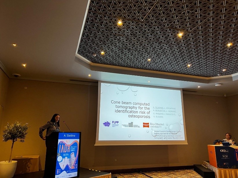
21.09.2023.- 23.09.2023.
Anda Slaidiņa presented a study at the 2023 Oral Health Research Congress in Greece. The research aimed to assess the correlation between mandibular cortical bone thickness, measured from cone-beam computed tomography (CBCT) images, and bone mineral density (BMD). It also evaluated whether CBCT images can be used to predict osteoporosis risk in postmenopausal women.
Reference:
Anda Slaidiņa, Laura Krumpāne, Anastasija Beibakova, Laura Neimane, Oskars Radziņš, Ivars Namatevs, Kaspars Sudars. “Cone Beam Computed Tomography for the Identification Risk of Osteoporosis.” Presented at the 2023 Oral Health Research Congress, a joint event of the Continental European Division (CED-IADR) and the Scandinavian Division (NOF) of the International Association for Dental Research, September 21–23, 2023, in Rhodes, Greece. Available online: https://iadr.abstractarchives.com/abstract/ced-iadr2023-3932484/cone-beam-computed-tomography-for-the-identification-risk-of-osteoporosis
22.09.2023.
A study aimed at developing a modular deep neural network for identifying osteoporosis risk based on 3D cone-beam computed tomography (CBCT) scans of the oral, facial, and maxillofacial region has been published in the Tomography journal, indexed by SCOPUS. The research also evaluated the network’s predictive capability for osteoporosis detection.
Reference:
Namatevs, I., Nikulins, A., Edelemers, E., Neimane, I., Slaidiņa, A., Radziņš, O., Sudars, K. “Modular Neural Networks for Osteoporosis Detection in Mandible Cone-Beam Computed Tomography Scans.” In Tomography, 2023, 9(5), 1772–1786.
Available online: https://doi.org/10.3390/tomography9050141
05.10.2023. – 06.10.2023.
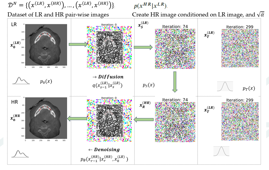
Namatevs I. participated in the 2023 IEEE 64th International Scientific Conference on Information Technology and Management Science at Riga Technical University (ITMS) with a presentation: Denoising Diffusion Algorithm for Single Image In-plaine Super-resolution in CBCT Scans of the Mandible, which was published later. The aim was to propose an accelerated algorithm for the super-resolution of medical images based on the denoising diffusion probability modeling approach.
Reference:
Namatevs, I., Sudars, K., Nikulins, A., Slaidiņa, A., Neimane, L., Radziņš, O., Edelemers, E. “Denoising Diffusion Algorithm for Single Image In-Plane Super-Resolution in CBCT Scans of the Mandible.” In 2023 IEEE 64th International Scientific Conference on Information Technology and Management Science of Riga Technical University (ITMS 2023), Riga, October 5, 2023, 194564.
Available online: 10.1109/ITMS59786.2023.10317791
13.11.2023.
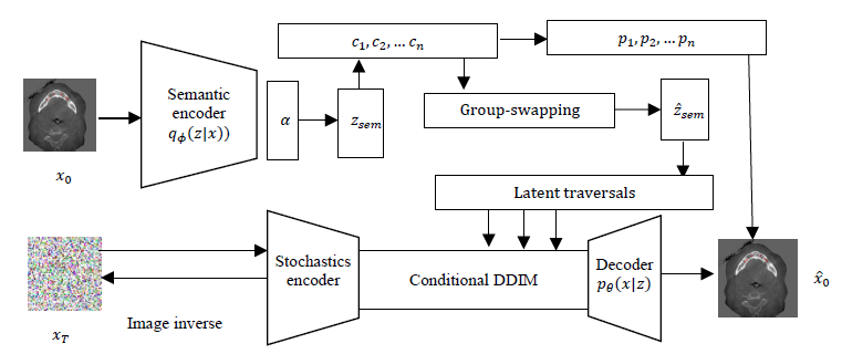
A research paper has been published, proposing the application of a conditional diffusion modeling approach with a Dirichlet variation autoencoder to disentangle and explain defined mandibular features in latent space.
Reference:
Namatēvs, I., Sudars, K., Ņikuļins, A., Slaidiņa, A., Neimane, L., Radziņš, O. “Towards Explainability of the Latent Space by Disentangled Representation Learning.” In Information Technology and Management Science, 2023(26), 41–48.
Available online: https://itms-journals.rtu.lv/article/view/itms-2023-0006
12.09.2024. – 14.09.2024.
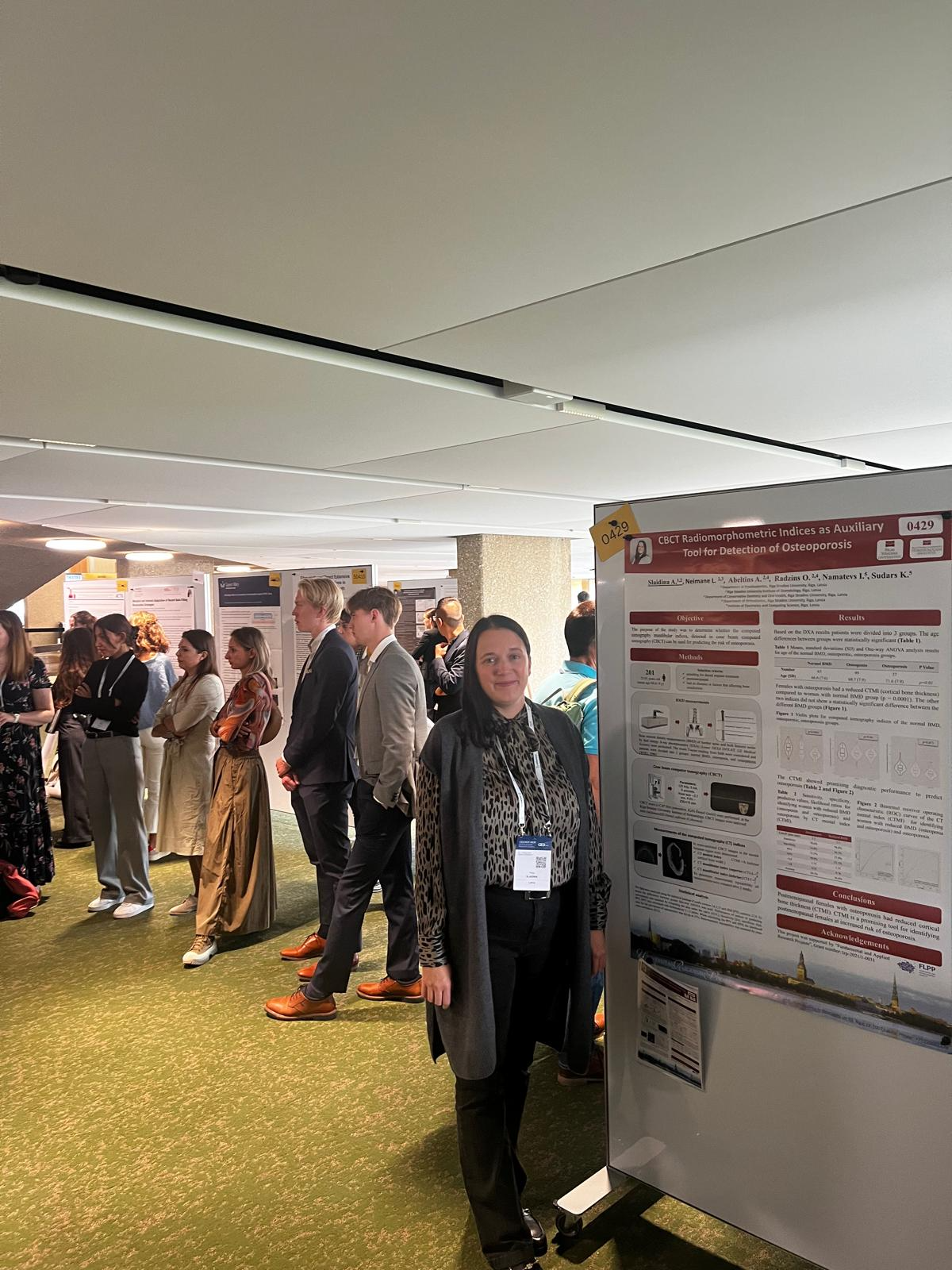
A. Slaidiņa participated with a poster presentation at the 2024 CED/NOF-IADR Oral Health Research Congress held in Geneva, Switzerland.
Reference:
Slaidiņa, A., Neimane, L., Ābeltiņš, A., Radziņš, O., Namatēvs, I., Sudars, K. “CBCT Radiomorphometric Indices as Auxiliary Tool for Detection of Osteoporosis.”
08.10.2024.
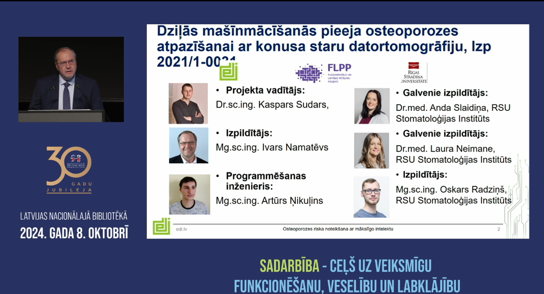
I. Namatēvs participated in the conference “Collaboration – The Path to Successful Functioning, Health, and Well-being,” organized by the rehabilitation center “Mēs esam līdzās” in cooperation with the National Centre for Education and the Riga City Council. He presented the results and technologies developed within the OSTAK project, focusing on Osteoporosis Risk Detection Using Artificial Intelligence.
Available online: https://www.visc.gov.lv/lv/media/27849/download?attachment
12.10.2024.
A. Slaidiņa participated in the EAO Congress with an e-poster presentation. The results of the study have been published in the SCOPUS-indexed (Q1) journal Clinical Oral Implants Research. The research examines the effect of osteoporosis on bone quantity and quality in the edentulous mandible.
Reference:
Slaidiņa, A., Neimane, L., Radziņš, O., Namatēvs, I., Sudars, K. “E-Poster: The Effect of Osteoporosis on the Bone Quantity and Quality of the Edentulous Mandible.” In Clinical Oral Implants Research, 2024, 35(28), 129–130, John Wiley & Sons.
Available online: https://doi.org/10.1111/clr.14366
24.10.2024. – 26.10.2024.
Slaidina A. participated in the 31st Annual Scientific Meeting of EAO and IAO, SIdP joint meeting 2024, Milan, Italy, 24-26 October 2024, and presented a joint research poster titled “The effect of osteoporosis on the bone quantity and quality of the edentulous mandible.”
19.12.2024.
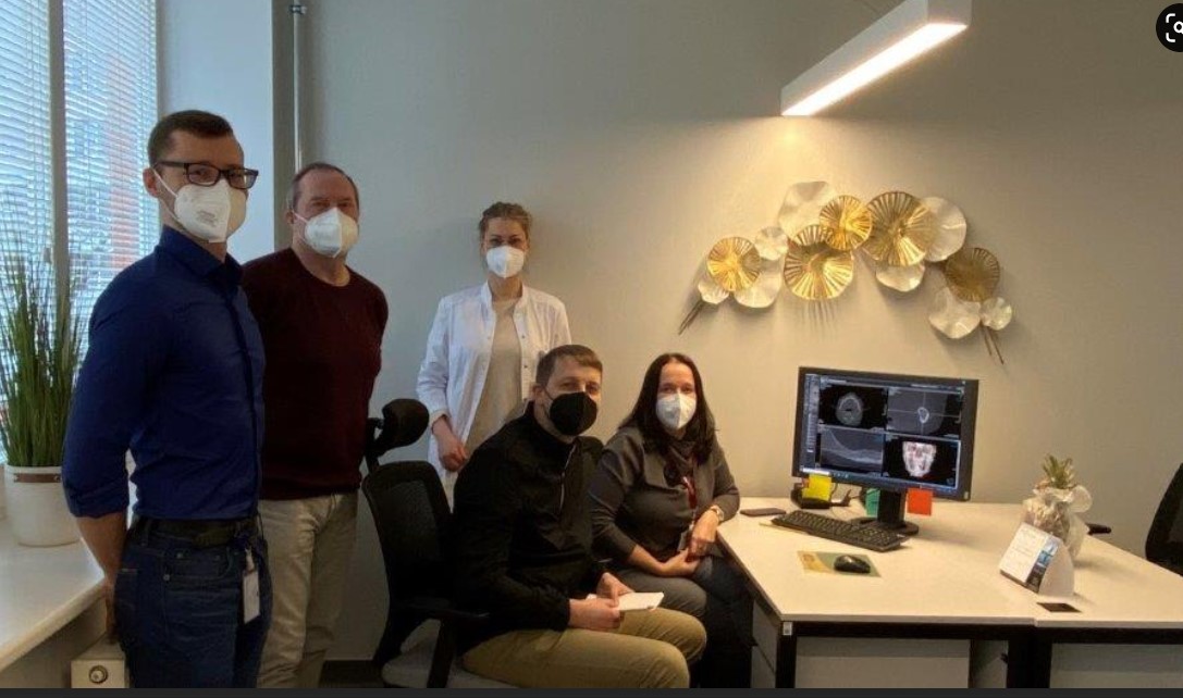
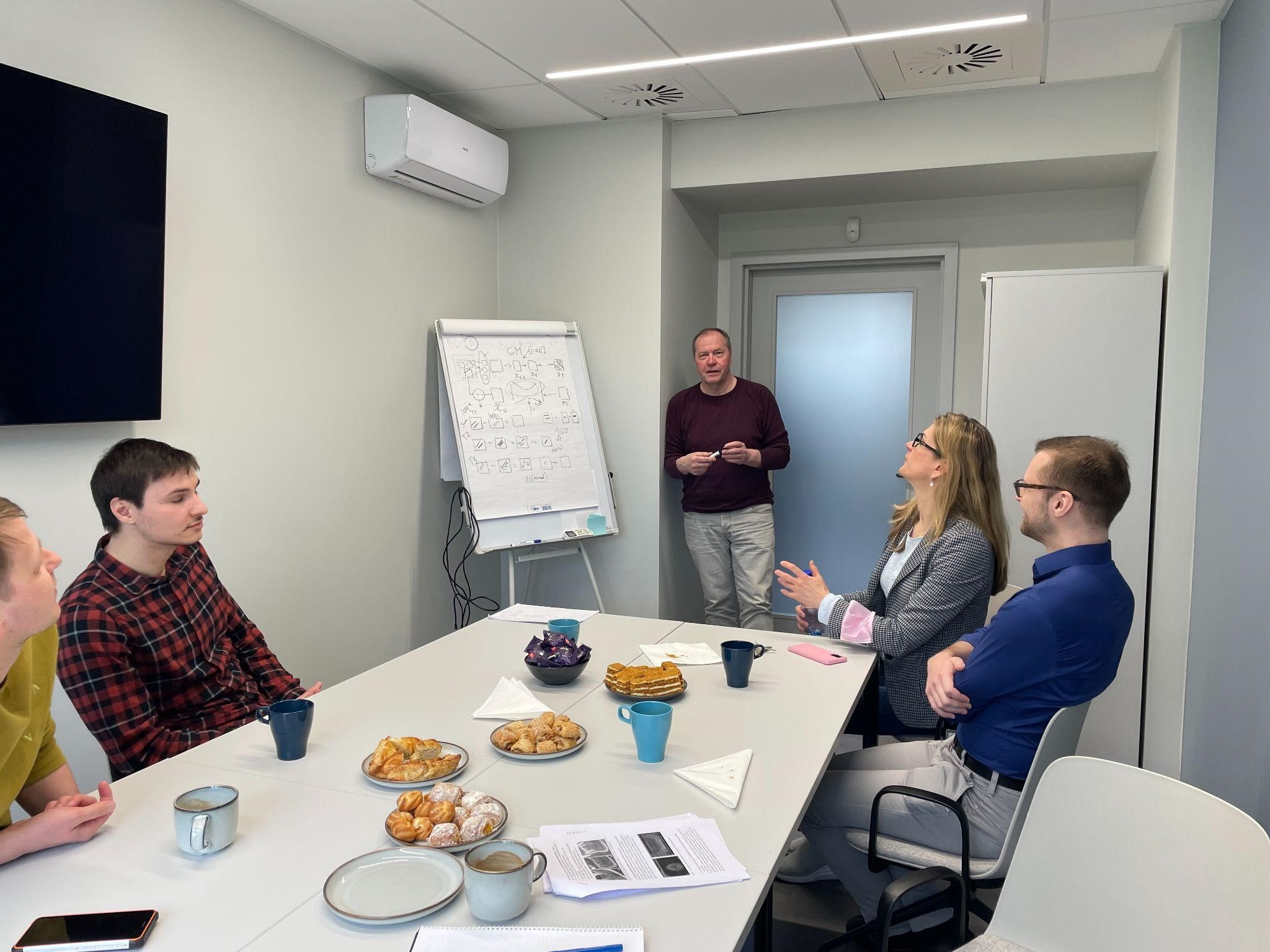
A final project meeting was held for researchers involved in the OSTAK project. The research results were summarized, and discussions focused on further developing the technologies, algorithms, and methods and potential future collaboration opportunities.
Summary of the OSTAK project:
Two new technologies were developed as part of the project:
- OSTAK osteoporosis detection. The three-stage modular DNN technology for detection
- OSTAK ViT Mandible Slice Detection.
A unique OSTAK dataset with 209 patients.
Three SCOPUS-indexed research papers and one international peer-recognised paper have been published.
Three post-graduate theses were written and defended as part of the project:
- Krumpāne Laura. Effect of reduced bone mineral density on the density. Defended on 18 January, 2023
- Baibakova Anasatasija. Effect of osteoporosis on the density of the jaw bones. Defended on 18 January, 2023.
- Emīlija Emija Ešenvalde-Krauze. Osteoporosis and Dental Implants. Defended on 7
January, 2024
- Two research proposals were submitted during the project period.
Participating scientists